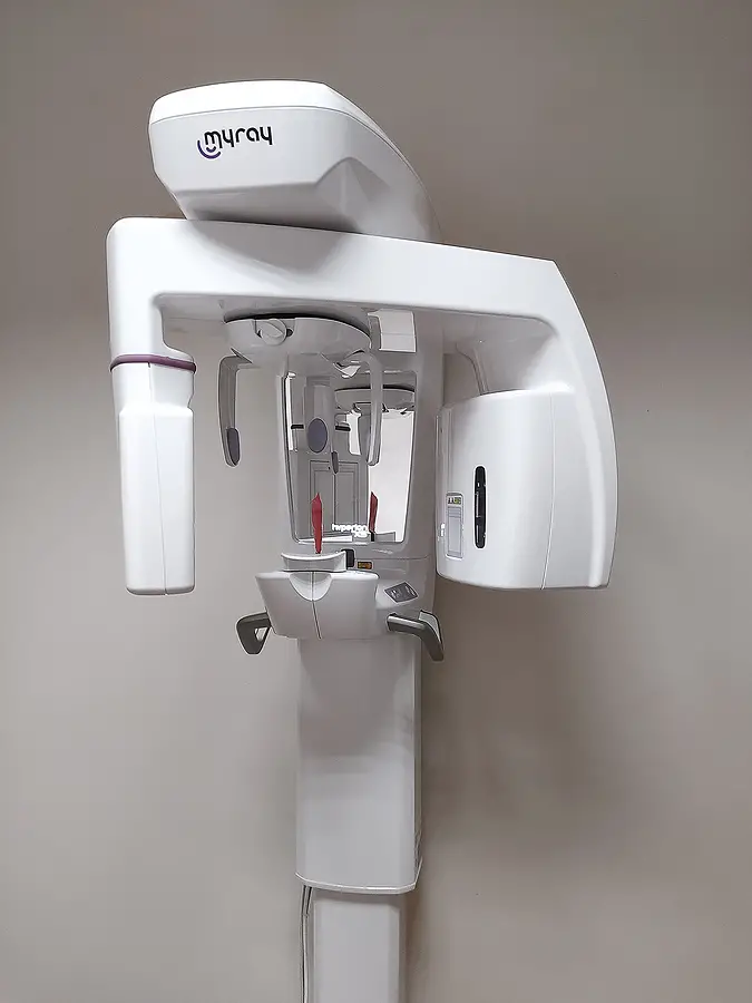
Cone Beam 3D Imaging is an advanced dental imaging technology that utilizes a cone-shaped beam to capture detailed, three-dimensional images of the teeth, jaws, and surrounding structures. Unlike traditional X-rays that produce two-dimensional images, Cone beam 3D imaging provides comprehensive views from multiple angles, offering dentists in Eagle Grove, IA, a more accurate and detailed dental and facial anatomy assessment. This technology is beneficial in various dental procedures, including implant placement, orthodontic treatment planning, and diagnosing complex dental issues, as it allows for precise measurements, enhanced visualization, and improved treatment outcomes while minimizing radiation exposure for patients.

How Is Cone Beam 3D Imaging Reshaping Dentistry?
Cone beam 3D imaging (CBCT) is revolutionizing dentistry by providing dentists with unparalleled diagnosis, treatment planning, and patient care capabilities. With its ability to produce detailed three-dimensional images of the teeth, jaws, and surrounding structures, CBCT offers a level of clarity and precision previously unattainable with traditional imaging methods. This advanced technology enables dentists at Moffitt Dental, to detect dental issues with greater accuracy, plan treatments more effectively, and collaborate seamlessly with interdisciplinary teams, ultimately leading to superior clinical outcomes and enhanced patient satisfaction. As CBCT continues to evolve, it promises to reshape the landscape of dentistry, driving innovation and advancements in patient care for years to come.
The Applications of Cone Beam 3D Imaging
Dental Implant Planning
Cone beam 3D imaging in Eagle Grove, IA, allows dentists to assess bone density, quality, and volume accurately, making it an invaluable tool for planning dental implant placement. By visualizing the jawbone in three dimensions, dentists can determine the optimal location and angle for implant placement, ensuring long-term stability and success.
Orthodontic Treatment Planning
Cone Beam 3D imaging provides orthodontists with comprehensive views of the teeth, jaws, and facial structures, enabling precise treatment planning for orthodontic interventions. This technology allows for assessing dental and skeletal relationships, identifying impacted teeth, and visualizing anatomical structures that may affect treatment outcomes. Contact us today!
Endodontic Diagnosis and Treatment
In endodontics, cone beam 3D imaging aids in the diagnosis and treatment planning of complex root canal cases. Dentists can visualize the tooth's internal anatomy, including the number and curvature of root canals, facilitating more accurate diagnosis and treatment planning. Additionally, cone beam 3D imaging can help identify the presence of periapical lesions or other pathology that may require intervention.
Temporomandibular Joint (TMJ) Assessment
Cone beam 3D imaging allows dentists to assess the temporomandibular joint (TMJ) and surrounding structures in three dimensions, aiding in diagnosing and treating TMJ disorders. Dentists can evaluate joint morphology, condylar position, and signs of arthritis or degenerative changes, helping to develop customized treatment plans for TMJ dysfunction.
Oral and Maxillofacial Surgery
Cone beam 3D imaging is widely used in oral and maxillofacial surgery for preoperative planning and assessment of complex anatomical structures. Surgeons can visualize the relationship between teeth, bones, nerves, and soft tissues, allowing for precise surgical planning and minimizing the risk of complications during procedures such as impacted tooth extraction, orthognathic surgery, and bone grafting.
The Benefits of Cone Beam 3D Imaging
Cone beam 3D imaging, also known as CBCT (Cone Beam Computed Tomography), provides many benefits in dentistry, revolutionizing the diagnostic and treatment planning process.
Firstly, CBCT provides detailed three-dimensional images of the teeth, jaws, and surrounding structures with exceptional clarity and precision. This comprehensive visualization allows dentists to accurately assess dental anatomy, identify pathology, and plan treatments with unparalleled accuracy. By capturing images from multiple angles, CBCT provides a comprehensive view of the patient's oral and maxillofacial anatomy, facilitating more informed decision-making and improving treatment outcomes.
Secondly, cone beam 3D imaging offers enhanced diagnostic capabilities, enabling dentists to detect dental issues that may not be visible on traditional two-dimensional X-rays. CBCT scans provide detailed information about bone density, volume, and morphology, aiding in diagnosing conditions such as dental caries, periodontal disease, and temporomandibular joint (TMJ) disorders.
Thirdly, CBCT plays a crucial role in treatment planning for various dental procedures, including dental implant placement, orthodontic treatment, and endodontic therapy. By providing precise measurements and visualization of anatomical structures, CBCT assists dentists in planning procedures with greater accuracy and predictability. This leads to more successful outcomes, reduced treatment times, and minimized patient risks. Additionally, CBCT enables dentists to simulate surgical procedures and anticipate potential complications, allowing for meticulous preoperative planning and optimization of treatment strategies.
Fourthly, cone beam 3D imaging enhances patient safety by minimizing radiation exposure compared to conventional CT scans. While CBCT utilizes a cone-shaped beam of radiation to capture images, it delivers lower levels of radiation than traditional CT scans, making it a safer option for patients.
Lastly, CBCT scans can be obtained quickly and conveniently in the dental office, eliminating referrals to external imaging centers and streamlining the diagnostic process. Overall, the benefits of cone beam 3D imaging in dentistry are vast, encompassing improved diagnostic accuracy, enhanced treatment planning, and increased patient safety, ultimately leading to superior clinical outcomes and patient satisfaction.
Unlock the future of dental diagnosis and treatment with cone beam 3D imaging. Visit Moffitt Dental at 322 S Commercial Ave, Eagle Grove, IA 50533, or call (515) 448-4852 to Discover the precision, accuracy, and comprehensive imaging capabilities of CBCT and transform your dental care experience.